
Art and Technology
Art and Technology
The third installment of a multi-platform series exploring the connections between art and the topics that move our world: climate, globalism, technology, science, health, law, finance, theft, international relations, destruction, religion and death.
The intersections between art and technology make scintillating headlines when cutting edge silicon valley tech is applied to the art world, giving a futuristic transfusion to a sector long-steeped in tradition. It seems a page has been ripped out of a sci-fi novel when blockchain is used to track provenance, synthetic DNA is added to works, and algorithms learn to distinguish the style of individual artists’ brushstrokes. These flashy examples barely scratch the surface of the deeply entrenched relationship between art and technology. Technology can facilitate the conservation, study, documentation, reconstruction, authentication and sale of art. Art and technology will explore a number of procedures including chemical analyses of physical samples, noninvasive diagnostics using advanced imaging techniques, and scanning and rendering methods for conservation.
Chemical Analysis of Physical Samples
Some of the most sophisticated technology used in the art world today requires something distinctly old school: a physical sample. These samples can be smaller than a single grain of sand and can provide information integral to a conservator’s work. When a conservator begins work on a painting, they must know the chemical makeup of the paint, canvas and any varnish applied to the piece. Typically, they deal with older works and for centuries, artists mixed their own paints. The composition of these paints can be simplified in two parts: the pigment and the binder. Binders are generally organic in nature- think egg yolk or oil- while pigments are typically mineral or organic- think ground parts of flora and fauna. It’s this compound that conservators sample for analysis. This analysis of paint can be handled by a two-step process known as gas chromatography and mass spectrometry. Chromatography, simply put, is the science of separation. It’s the process through which the various binders and different pigments are isolated. The next step, Mass Spectrometry, analyzes the mass-to-charge ratio of ions to tell us what is present in the sample. The data will usually be presented as a vertical bar graph, and will distinguish each ion present as well as its relative abundance. Conservators can correlate these results with graphs showing known ions to determine what mineral or organic materials the artist used to create the paint and varnish.
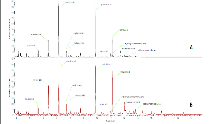
Total ion current chromatogram of A) a white paint sample and B) a dark-brown paint sample from "Light" panel and after transesterification and GC-MS analysis (internal standard = nonadecanoic acid)

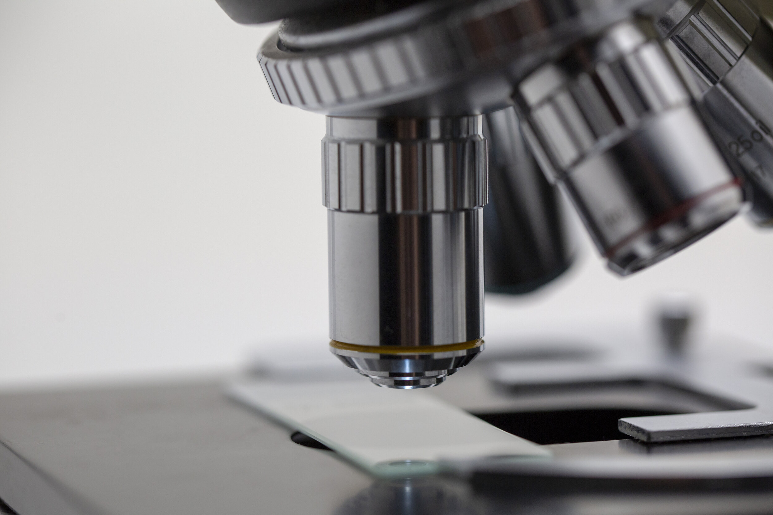
Close up of a Microscope
In addition to chemical makeup, physical samples from artworks can also provide information on the age of a work through a process known as radiocarbon dating. When lead in paint oxidizes, it forms a carbonate, capturing a snapshot of the radioactive carbon-14 concentration in the atmosphere at the time the artwork was created. The level of carbon-14 measured can be converted into an actual year by consulting a chart of historical atmospheric concentrations. For paintings made since the mid twentieth century, it’s possible to be accurate within a year or two due to the rapid change in CO2 levels. Conservators can also use physical samples to date a painting through optical microscopy: analyzing paint samples under a microscope. By viewing the pigments at hundreds of times the magnification, a trained eye can distinguish pigments which may have been unavailable to artists before certain time periods in history, helping to verify the authenticity of a work or properly conserve it.
Advanced Imaging Technology
The tech used in the art world, however, isn’t limited to the analysis of physical samples. Researchers can answer many questions about a work and its artistic process without ever laying a hand on the work itself. They can discover details about any past restoration processes, how close the final work was to the sketches underneath the pigments, and if the canvas was previously used for another work. This information can be uncovered without any damage to the work through advanced imaging technology, making use of the electromagnetic spectrum. When we look at a work we only see it across the visible light portion of the spectrum.

Electromagnetic Spectrum

Pablo Picasso, Woman Ironing. The visual light view (left) and the infrared reflectography image.
When we view the works through technology designed to focus on parts of the spectrum beyond visible light, we reveal layers of the works concealed to the naked eye. This is called multispectral imaging and includes infrared reflectography, macro x-ray fluorescence and ultraviolet imaging, each revealing different layers. For example, there is a near-infrared range where the wavelengths are longer than those of visible light and are usually invisible. When using techniques to view the works on this portion of the spectrum, the pigments usually visible in paint now become invisible, revealing the underlying details of a painting. In a particularly stunning discovery using this infrared imaging technique, the conservation team at the Guggenheim found a long lost portrait hiding under Picasso’s painting, Woman Ironing.
Digital Rendering Techniques
When works are at risk of degradation or are too delicate to handle or display, conservators and preservationists can go one step further than investigative imaging and actually create digital renderings. These renderings can serve as heritage documentation, creating a snapshot in time of a work at risk, or as a blueprint for accurate reconstruction. Many forms of technology have been developed to aid in creating digital reproductions and virtual renderings of art and artifacts. Each technology is particularly suited to a different need. One can determine which technology to use based on the size and shape of the piece being rendered and the accuracy needed in the final product. Generally, more accuracy is needed for renderings used in restoration or heritage documentation while less precision is necessary in renderings used for study or digital access.

Point cloud' of the Three Graces by Antonio Canova © Factum Foundation
3D models used for study and access are often produced through photogrammetry. Photogrammetry is a technique that constructs 3D measurements based on a set of overlapping images taken around the object from different angles. Without additional handling of the work, the captured images can be combined into a crude rendering. The more uniform the lighting, and the higher the quality of the images, the more accurately represented the proportions of the object. The more precise and systemic the process of capturing the photos, the more clear and complete the rendering.
The cultural sector also makes use of laser scanners for precision renderings used in restoration or heritage documentation. Terrestrial laser scanners measure distances employing a laser ranging system using one of two methods: pulsed-based or phase-based measurements. Each has its own advantages. A pulse-based system sends out a laser beam and waits for its return before sending out another beam. It collects points one by one with incredible accuracy and range, but relatively slowly. A phase-based laser sends out continuous beams and sees a decrease in accuracy over a larger range. It also requires an incredible amount of power to operate. With either approach, the distance measurements are combined with angular measurements to produce 3D information in the form of points, generating a point cloud. Once the point cloud is created, a colorless rendering of the cultural asset will be produced (see image). This can be easily colored to look like the real work using photographs as a reference.

Point cloud Rendering
a Simplified schematic layout of the multi-scale spectral domain OCT system (top view). b setup in lab conditions. c MS-OCT probe in front of the painting.
When dealing with larger assets such as cultural artifacts or structures, terrestrial scanners are best. Yet, there are other specialty scanners which focus on more refined details such as the delicate topography on the surface of a painting. Requiring the highest level of accuracy and precision, incredibly subtle details of impasto (varying paint thickness) and cracks developed over time by two techniques: 3D digital microscopy and MS-OCT (multi-scale optical coherence tomography.) MS-OCT operates by dividing the artwork into sections, like tiles on a floor. Each “tile” is scanned using sensitive scanners which map the surface of the area and then one large pass is taken over the full artwork to assist the algorithm in stitching the tiles together.

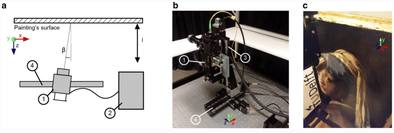
Fringe-encoded stereo imaging a denotes the cameras, b the projector, c the vertical stage(s) and d/e the horizontal stage, ββ the imaging angle, αα the tilt angle of the lenses and l the working distance to the painting
With fringe-encoded stereo imaging, there are two scanning systems, fitted on either side of a projector opposite the painting’s surface. 3D digital microscopy uses the principle of focus stacking for 3D imaging, by employing a microscope suspended over the work at a magnification of 140× and using a motorized stand to scan the entire work from the two angles simultaneously at varying focus layers. These are then combined into one single all-in-focus image referred to as multi-focus or extended depth-of-field. It is through these techniques that conservationists have studied the surface of Vermeer’s, Girl with a Pearl Earring. Conservationists have gained unparalleled insight into surface features, specifically related to ‘moating’ around impasto, the effects of paint consolidation in earlier restoration campaigns and aging, through visualisation of the pattern of the cracks.

Existing documentation visualising the surface topography of ‘Girl with a Pearl Earring’. a Raking light photography is traditionally used to emphasise the topographical variations of the surface, visualising cracks and other unevenness of the surface (Courtesy of René Gerritsen Art & Research Photography). b The crack pattern of the painting was documented by manually tracing the cracks onto a transparent polyester film, placed on the painting (Courtesy of Jørgen Wadum [4]).
The red inserts show an enlargement of the area around the pearl earring.
Whether the goal is to facilitate access, documentation, study, or conservation, technology has enabled the art world to become more precise and efficient in how we handle works. Technology in art plays an indispensable role in the protection and care of collections, regardless of the field in which it originated. As technology continues to develop, we will, no doubt, reveal more secrets hidden in the art we’ve appreciated for centuries. We will also surely learn to care more efficiently for our masterpieces, keeping them true to their original aesthetic for generations to come.
Subscribe to Art and
Sign up and get the next installment sent to your email.
Sources
Brooks, Kevin R. “Depth Perception and the History of Three-Dimensional Art: Who Produced the First Stereoscopic Images?” I-Perception, vol. 8, no. 1, Jan. 2017. PubMed Central, doi:10.1177/2041669516680114.
Candeias, Antonio, and Juan Manuel Madariaga. “Applications of Raman Spectroscopy in Art and Archaeology.” Journal of Raman Spectroscopy, vol. 50, no. 2, 2019, pp. 137–42. Wiley Online Library, doi:10.1002/jrs.5571.
Carbon, Claus-Christian, and Vera M. Hesslinger. “DaVinci’s Mona Lisa Entering the next Dimension.” Perception, vol. 42, no. 8, 2013, pp. 887–93. PubMed, doi:10.1068/p7524.
Chun, Rene. “These Four Technologies May Finally Put an End to Art Forgery.” Artsy, 18 July 2016, https://www.artsy.net/article/artsy-editorial-these-four-technologies-may-finally-put-an-end-to-art-forgery.
Davis, Annabelle, et al. “Pilbara Rock Art: Laser Scanning, Photogrammetry and 3D Photographic Reconstruction as Heritage Management Tools.” Heritage Science, vol. 5, no. 1, July 2017, p. 25. BioMed Central, doi:10.1186/s40494-017-0140-7.
Hendriks, Laura, et al. “The Ins and Outs of 14C Dating Lead White Paint for Artworks Application.” Analytical Chemistry, vol. 92, no. 11, June 2020, pp. 7674–82. ACS Publications, doi:10.1021/acs.analchem.0c00530.
“How Artists’ Paints Are Made.” The Economist. The Economist, https://www.economist.com/the-economist-explains/2014/06/23/how-artists-paints-are-made. Accessed 6 Oct. 2020.
“Infrared Reflectography in Art Analysis, Conservation & History.” IRINFO.Org, https://irinfo.org/articles-2019/infrared-reflectography-5-1-19-tucker/. Accessed 4 Oct. 2020.
Sanyova, Jana, et al. “Unexpected Materials in a Rembrandt Painting Characterized by High Spatial Resolution Cluster-TOF-SIMS Imaging.” Analytical Chemistry, vol. 83, no. 3, Feb. 2011, pp. 753–60. ACS Publications, doi:10.1021/ac1017748.
Saverwyns, Steven, et al. “Macro X-Ray Fluorescence Scanning (MA-XRF) as Tool in the Authentication of Paintings.” Microchemical Journal, vol. 137, Mar. 2018, pp. 139–47. ScienceDirect, doi:10.1016/j.microc.2017.10.008.
Serrano, Ana, et al. “Investigation of Crimson-Dyed Fibres for a New Approach on the Characterization of Cochineal and Kermes Dyes in Historical Textiles.” Analytica Chimica Acta, vol. 897, Oct. 2015, pp. 116–27. DOI.org (Crossref), doi:10.1016/j.aca.2015.09.046.
“The Microscope in Art Conservation and Authentication Studies.” McCrone, 9 Nov. 2003, https://www.mccrone.com/mm/the-microscope-in-art-conservation-and-authentication-studies/.
“Time-of-Flight vs. Phase-Based Laser Scanners: Right Tool for the Job.” SPAR 3D, 22 June 2004, https://www.spar3d.com/news/related-new-technologies/time-of-flight-vs-phase-based-laser-scanners-right-tool-for-the-job/.
“---.” SPAR 3D, 22 June 2004, https://www.spar3d.com/news/related-new-technologies/time-of-flight-vs-phase-based-laser-scanners-right-tool-for-the-job/.
Today, Chromatography. “Uncovering Art’s Mysteries with Chromatography.” Chromatography Today, https://www.chromatographytoday.com/news/hplc-uhplc/31/breaking-news/uncovering-artrsquos-mysteries-with-chromatography/33105. Accessed 5 Oct. 2020.